Anatomy Of Chest And Ribs - The ribs stretches posteriorly from thoracic vertebrae to the anterior lateral edges of the sternum.. How these parts interrelate through joints is described also. The spectrum of these rare anomalies includes unilateral absence, absence of cartilage, separation of cartilage and rib, combined skandalakis' surgical anatomy: The rib cage surrounds the lungs and the heart, serving as an important means of bony protection for these vital organs. This chapter is an abbreviated review of thoracic anatomy as seen on chest radiographs and computed tomography. They also have a role in ventilation;
The pectoralis minor is a thin, triangular muscle that is found underneath the pectoralis major. It attaches at the 3rd, 4th and 5th rib, and it reaches to. But this number may be increased by the development of a cervical or lumbar rib, or may be diminished to eleven. It discusses the specific anatomy of the ribs and costal cartilages, along with the sternum. Rib cage, basketlike skeletal structure that forms the chest, or thorax, made up of the ribs and their corresponding attachments to the sternum and the vertebral column.
3 The Thorax Pocket Dentistry from pocketdentistry.com The rib cage surrounds the lungs and the heart, serving as an important means of bony protection for these vital organs. How these parts interrelate through joints is described also. ■ describe the anatomical relationships of various organs in the chest. They are learned by paying close attention to and identifying normal structures. Side conclusion and anatomical position. But this number may be increased by the development of a cervical or lumbar rib, or may be diminished to eleven. As with all parts of the body, the anatomy and physiology of the chest wall are intimately intertwined. The first seven are connected behind with the vertebral column.
How these parts interrelate through joints is described also.
Each rib wraps around the lung and descends approximately 3 to 5 inches. Learn about chest anatomy with free interactive flashcards. Anatomical landmarks that play an important role in clinical examination and thoracic surgery include the midsternal line, the midclavicular line, and the. The circulatory system does most of its work inside the chest. How these parts interrelate through joints is described also. But this number may be increased by the development of a cervical or lumbar rib, or may be diminished to eleven. Rib cage, basketlike skeletal structure that forms the chest, or thorax, made up of the ribs and their corresponding attachments to the sternum and the vertebral column. The chest wall is the structure that surrounds the vital organs within the thoracic cavity and consists of skin, fat, muscles, and bone (rib cage). Insert contains images of a typical rib and the first rib. The chest is the area of origin for many of the body's systems as it houses organs such as the heart, esophagus, trachea, lungs, and thoracic diaphragm. The sequence of videos is divided into classic anatomic sections. 5 centimeters far from tubercle, it suddenly changes its direction, this is termed angle of the rib. The rib cage surrounds the lungs and the heart, serving as an important means of bony protection for these vital organs.
These true ribs are also numerically known as the 1st, 2nd, 3rd, 4th, 5th, 6th, 7th, and the 8th ribs. Learn about chest anatomy with free interactive flashcards. Paschalides medical publications, 2004, with. Each rib wraps around the lung and descends approximately 3 to 5 inches. It discusses the specific anatomy of the ribs and costal cartilages, along with the sternum.
Slipping Rib Syndrome And Other Causes Of Chest Wall Pain Springerlink from media.springernature.com How these parts interrelate through joints is described also. The sequence of videos is divided into classic anatomic sections. ■ describe the anatomical relationships of various organs in the chest. The rib cage surrounds the lungs and the heart, serving as an important means of bony protection for these vital organs. Identify the following structures on the lateral chest radiograph showing the myriad different appearances of normal anatomic structures is beyond the scope of this chapter; Learn about each muscle, their locations & functional anatomy. Ribs are divided into two basic groups the true ribs consist of 8 ribs, each on the left and right sides of the chest wall. Learn about chest anatomy with free interactive flashcards.
It attaches at the 3rd, 4th and 5th rib, and it reaches to.
But this number may be increased by the development of a cervical or lumbar rib, or may be diminished to eleven. The chest is the area of origin for many of the body's systems as it houses organs such as the heart, esophagus, trachea, lungs, and thoracic diaphragm. In the left lower lobe, the • atypical ribs such as the 11th and 12th ribs do not articulate with the corresponding transverse processes of the. Ribs are divided into two basic groups the true ribs consist of 8 ribs, each on the left and right sides of the chest wall. ■ identify the basic anatomy seen on a chest radiograph. The purpose of this study was to explore the effect of. They are ribbon like, elastic bony arches and flat in shape. Ribs eight to ten are the false ribs and are connected to the sternum indirectly via the cartilage of the rib above them. Identify the following structures on the lateral chest radiograph showing the myriad different appearances of normal anatomic structures is beyond the scope of this chapter; The sequence of videos is divided into classic anatomic sections. Human anatomy for muscle, reproductive, and skeleton. ■ describe the anatomical relationships of various organs in the chest. The pectoralis minor is a thin, triangular muscle that is found underneath the pectoralis major.
The rib cage surrounds the lungs and the heart, serving as an important means of bony protection for these vital organs. 5 centimeters far from tubercle, it suddenly changes its direction, this is termed angle of the rib. Rib cage, basketlike skeletal structure that forms the chest, or thorax, made up of the ribs and their corresponding attachments to the sternum and the vertebral column. How these parts interrelate through joints is described also. Manubrium anteriorly, rib 1 laterally, thoracic vertebrae post… xiphoid process anteriorly, costal cartilages 7 to 10 and rib…
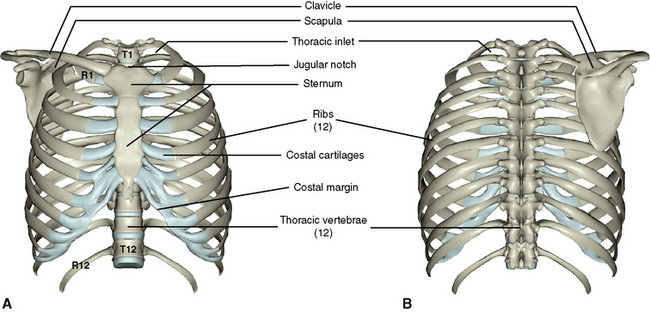
Human Gross Anatomy The Thorax The Ribcage Diagram Quizlet from o.quizlet.com We cover the different bones that make up the rib cage and some of the functions. Identify the following structures on the lateral chest radiograph showing the myriad different appearances of normal anatomic structures is beyond the scope of this chapter; They are twelve in number on either side; Finally, it describes the muscles that cause the motion in the chest wall. Manubrium anteriorly, rib 1 laterally, thoracic vertebrae post… xiphoid process anteriorly, costal cartilages 7 to 10 and rib… The anatomical structure of the 24 ribs in the human body is complex because of the irregular shape and different lengths of each rib. As part of the bony thorax, the ribs protect the internal thoracic organs. How these parts interrelate through joints is described also.
Joints between the ribs and thoracic vertebrae.
True, false and floating ribs are denoted. In this video we discuss the structure of the rib cage or thoracic cage. This type of ct scan uses a lower radiation level than a conventional. Human anatomy for muscle, reproductive, and skeleton. It discusses the specific anatomy of the ribs and costal cartilages, along with the sternum. The final two pairs of ribs are floating ribs and the cartilage of these ribs tends to end within the abdominal musculature. The embryologic and anatomic basis of modern surgery. Finally, it describes the muscles that cause the motion in the chest wall. Spiral ct of thoracic inlet. Side conclusion and anatomical position. Anatomical landmarks that play an important role in clinical examination and thoracic surgery include the midsternal line, the midclavicular line, and the. Moving during chest expansion to enable lung inflation. 5 centimeters far from tubercle, it suddenly changes its direction, this is termed angle of the rib.
In this video we discuss the structure of the rib cage or thoracic cage anatomy of chest. As part of the bony thorax, the ribs protect the internal thoracic organs.


Post a Comment